多重离子束成像
Multiplexed Ion Beam Imaging (MIBI)
组织微环境的综合空间分析
Comprehensive spatial analysis of the tissue microenvironment
多路离子束成像 (MIBI™) 技术实现了高清空间蛋白质组学,该技术独特地提供了对组织微环境的可操作分析,为疾病状态、作用机制和患者反应提供了新的见解。
|
|
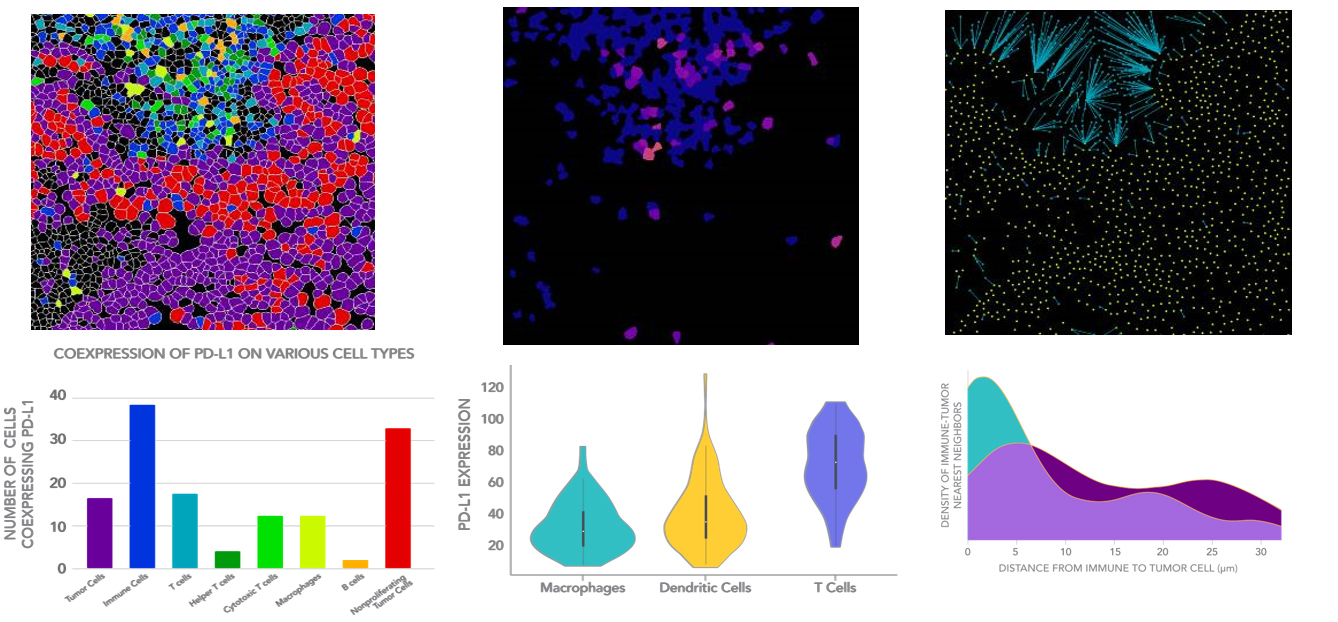
CELL CLASSIFICATION
Identify and enumerate cell populations
Visualize the tissue microenvironment |
PROTEIN QUANTIFICATION
Quantify expression of checkpoints and other key proteins with spatial context |
|
SPATIAL ANALYSIS
Analyze interactions between target and effector cells
Map tumor-immune boundaries
|
|
|
可以为治疗选择提供信息的高清空间蛋白质组学 Gain deeper understanding of the tumor microenvironment and reveal actionable insights. Identify responder populations or predict patient response. Achieve quantitative single-cell phenotype mapping at subcellular resolution and identify key mechanisms of action with clinical grade repeatability
深入了解肿瘤微环境并揭示可行的见解。 识别响应人群或预测患者响应。 以亚细胞分辨率实现定量单细胞表型映射,并确定具有临床级可重复性的关键作用机制。 |
MIBIscope™ System
|
Transforming Multiplexed Tissue Imaging |
改变多重组织成像 |
|
Visualize 40+ markers in a single scan |
在一次扫描中可视化 40 多个标记 |
|
Comprehensively phenotype immune
infiltrate |
综合表型免疫浸润 |
|
Quantify protein expression |
量化蛋白质表达 |
|
Profile tissue architecture |
轮廓组织结构 |
|
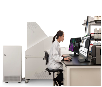
A revolutionary technology for analysis of the tumor microenvironment The MIBIscope™ System is a revolutionary imaging platform, enabling comprehensive phenotypic profiling and spatial analysis of the tissue microenvironment. The MIBIscope allows researchers to visualize over 40 markers simultaneously with higher sensitivity, resolution, and throughput than existing methods. 肿瘤微环境分析的革命性技术 MIBIscope™ 系统是一个革命性的成像平台,能够对组织微环境进行全面的表型分析和空间分析。 MIBIscope 允许研究人员同时可视化 40 多个标记,其灵敏度、分辨率和通量比现有方法更高。 |
|
|
|
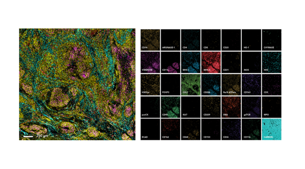
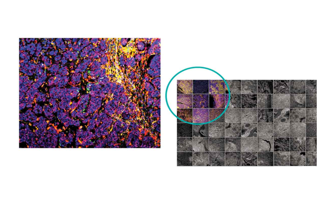
Visualize 40+ Markers in a Single Image 可视化40+标记在一个单一的图像 The MIBIscope enables over 40 markers to be visualized simultaneously with single step staining and single step imaging. MIBIscope images from a 3mm x 3mm scan of a granulomatous lung section from a Mycoplasm tuberculosis infected patient. The image to the left shows immune infiltrate in the infected tissue (CD45, yellow; CD31, red; SMA, blue; CD68, magenta). Zoomed in single-channel images shown to the right. MIBIscope 可以通过单步染色和单步成像同时显示 40 多个标记。 MIBIscope 图像来自对感染支原体的患者的肉芽肿肺切片进行 3mm x 3mm 扫描。 左图显示受感染组织中的免疫浸润(CD45,黄色;CD31,红色;SMA,蓝色;CD68,洋红色)。 放大的单通道图像如右图所示。 |
|
High Throughput Image up to 90 800×800 µm2 ROIs per day The MIBIscope has the reliability to run 24/7, allowing researchers to conduct studies on large clinical cohorts and image hundreds of samples a week. 每天成像多达 90 800×800 µm2 ROI MIBIscope 具有 24/7 全天候运行的可靠性,使研究人员能够对大型临床队列进行研究,并每周对数百个样本进行成像。 |
 |
|
 |
||
|
High Resolution
高分辨率 Achieve confocal resolution 实现共焦分辨率 The MIBIscope can be adjusted like an optical microscope with resolution settings from 350 nm to 1 µm. At higher magnifications the MIBIscope provides comparable resolution to confocal microscopy. MIBIscope 可以像光学显微镜一样进行调整,分辨率设置为 350 nm 至 1 µm。 在更高的放大倍率下,MIBIscope 可提供与共聚焦显微镜相当的分辨率。 |
||
 |
||
|
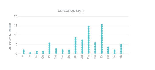
High Sensitivity 高灵敏度 Single molecule sensitivity 单分子灵敏度 The MIBIscope uses highly sensitive Secondary Ion Mass Spectrometry, allowing for single molecule detection as demonstrated in Science Advances and other recent publications. MIBIscope 使用高度灵敏的二次离子质谱法,允许进行单分子检测,如《Science Advances》和其他近期出版物中所示。 |
||
|
SPECIFICATIONS 规格
|
|
|
Available Biomarker Channels可用的生物标志物渠道 |
40 |
|
Resolution分辨率 |
350 nm – 1 µm |
|
ROI Size 视野 |
400x400 – 800x800 µm2 |
|
Acquisition Time 采集时间 |
(per 800x800 µm2 |
|
Coarse Resolution (1 µm)粗分辨率 |
35 min |
|
Fine Resolution (500 nm)精细分辨率 |
68 min |
|
Super-fine Resolution (350 nm) 超精细分辨率 |
139 min |
|
Lower Limit of Detection (number of Ab)检测下限 |
1 (113In) – 16 (166Er) |
|
Dynamic Range动态范围 |
5 log |
|
File Type文件类型 |
TIFF |
